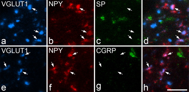
Figure 6.
VGLUT1, NPY, substance P (SP), and CGRP immunolabeling in boutons in lamina III of the ipsilateral lumbar spinal cord after unilateral L5 spinal nerve ligation. a–d show a single field scanned for VGLUT1, NPY, and substance P. NPY and VGLUT1 are colocalized in several boutons (some indicated with arrows), which belong to injured myelinated afferents. However, substance P immunoreactivity is not present in any of these (c, d). e–h show another field scanned for VGLUT1, NPY, and CGRP. Again, NPY and VGLUT1 are colocalized in certain boutons (some indicated with arrows), but these do not contain CGRP immunoreactivity (g, h). Images were obtained from stacks of five (a–d) or three (e–h) optical sections at 0.5 μm z-separation. Scale bar, 5 μm.

