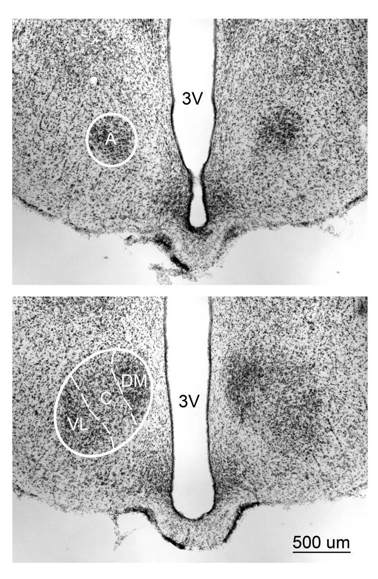
Figure 1.
Thionin-stained 40µm coronal section taken at two different rostrocaudal levels of the VMH of an adult WT male. (Top) Rostral most appearance of the anterior portion (A) (Bottom) Further caudally in the same material showing the Dorsomedial, (DM) Central (C), and the Ventrolateral (VL) portions of the VMH appear. The dashed lines separate the divisions. 3V = Third ventricle.

