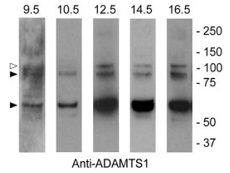
Fig. 2.

Immunoblot analysis of ADAMTS-1 in the developing mouse heart. ADAMTS-1 was detected by immunoblotting in whole embryonic heart extracts using guinea pig anti mouse ADAMTS-1 IgG. The open arrowhead points to the 110-kDa proform. Filled arrowheads point to the 87- and 65-kDa catalytically active forms. Extracts were electrophoresed under reducing conditions.
