
Fig. 6.
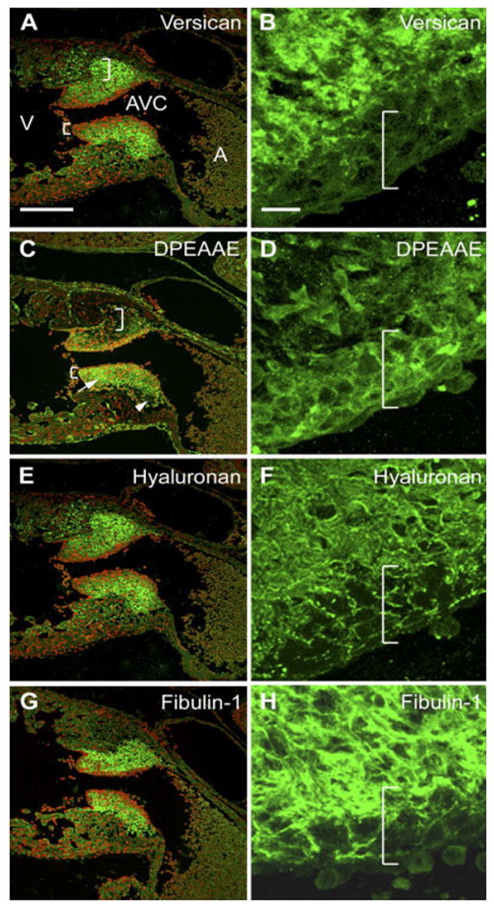
Localization of versican, its anti-DPEAAE–reactive cleavage product as well as fibulin-1 and hyaluronan in the 12.5 days post coitum (dpc) AVC cushion. A–H: Embryonic mouse heart serial sections (12.5 dpc) were immunolabeled with anti-versican GAGβ IgG (A,B), anti-DPEAAE IgG (C,D), bHABP (to detect HA; E,F), and anti–fibulin-1 IgG (G,H). A,C,E,G: Atrioventricular canal (AVC) regions. B,D,F,H: Enlargements of regions of inferior AVC cushions shown in A, C, E, and G, respectively (with the red channel turned off). Right brackets indicate regions of loosely packed mesenchyme within inferior AVC cushions. Left brackets indicate compacted cells subjacent to the endocardium. The arrow in D denotes anti-DPEAAE immunolabeling surrounding loosely packed mesenchymal cells of superior cushion that begins to appear at this stage. The arrowhead in D highlights deeper regions of the superior AVC cushion devoid of DPEAAE immunoreactivity but positive for anti-GAGβ reactive versican. V, ventricle; A, atrium. Nuclei are stained red using propidium iodide. Scale bars = 150 μm in A (applies to A,C,E,G), 15 μm in B (applies to B,D,F,H).