
Fig. 7.
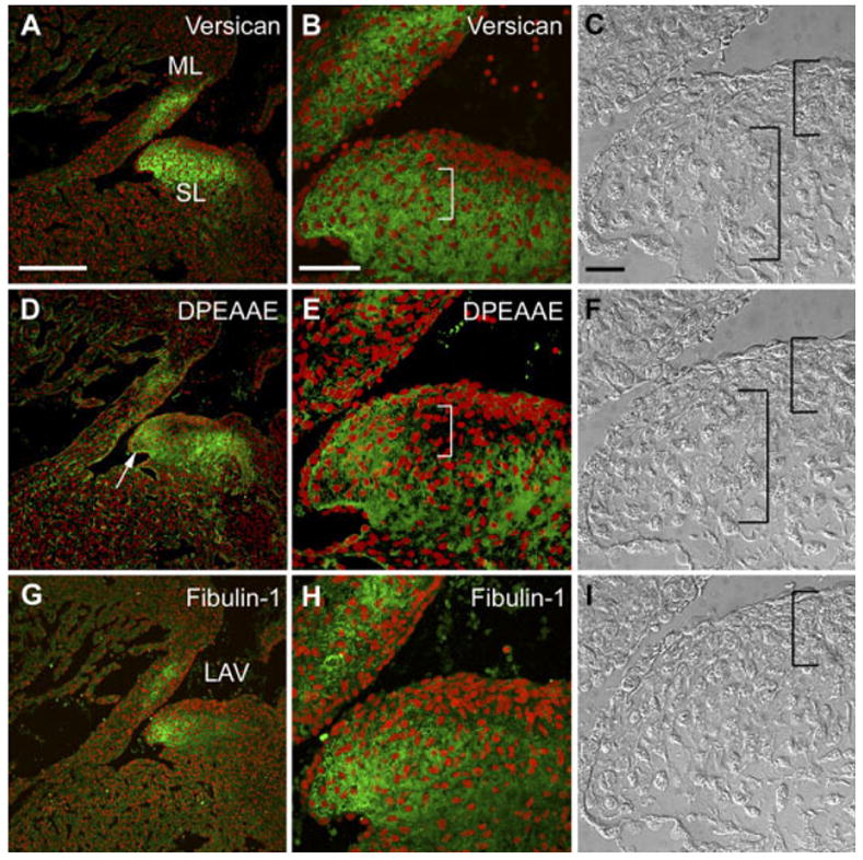
Localization of versican, its anti-DPEAAE–reactive cleavage product as well as fibulin-1 in the 14.5 days post coitum (dpc) atrioventricular canal (AVC) cushion. A,B,D,E,G,H: Embryonic mouse heart sections (14.5 dpc) were immunolabeled with anti-versican GAGβ IgG (A,B), anti-DPEAAE IgG (D,E) and anti-fibulin-1 IgG (G,H). B,E,H: Enlargements of regions of AVC cushions shown in A, D, and G, respectively. C,F,I: Nomarski differential interference contrast images corresponding to the immunostained sections shown in B, E, and H, respectively. Right brackets indicate regions of loosely packed mesenchyme within AVC cushions. Left brackets indicate compacted cells subjacent to the endocardium. ML, mural leaflet of the left AV region (LAV). SL, septal leaflet of the LAV. Arrow in E shows staining of the DPEAAE antibody subjacent to the endocardium of the elongating leaflet. Scale bars = 150 μm in A (applies to A,D,G), 50 μm in B (applies to B,E,H), 20 μm in C (applies to C,F,I).