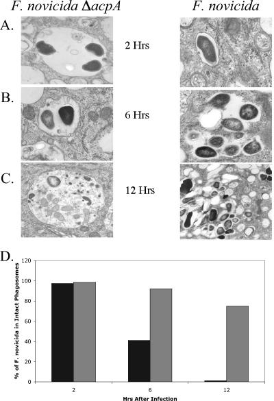
FIG. 4.
Transmission electron microscopy images of THP-1 cells infected with the acpA null strain of F. novicida (left panel) and the WT (right panel) obtained 2 h (A), 6 h (B), and 12 h (C) postinfection and (D) semiquantitative assessment of bacteria within/outside of phagosomes as determined by counting phagosomes in a minimum of 100 cross-sections/test group. Gray bars, ΔacpA; black bars, parental strain. The width of each panel in A, B, and C is 2.87 μm.

