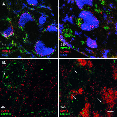
FIG. 1.
Migration of macrophage subsets in the spleen after infection with L. monocytogenes. Cryosections from the spleens of infected mice at the indicated time points were prepared, stained with appropriate fluorescent Abs, and analyzed using confocal microscopy. A. Clustering of ERTR-9+ marginal zone macrophages (green) around infection foci and migration of MOMA-1+ metallophilic marginal zone macrophages (red) into B-cell follicles (blue). B. Clustering of CD11b+ cells during infection with L. monocytogenes. CD11b+ cells are stained red, and laminin is green. At 4 h p.i., CD11b+ cells are scattered through the red pulp. At 24 h p.i., these cells form clusters around infection foci in the MZ area. No clusters were observed in the white pulp or surrounding the central arteriole. Arrows indicate central arterioles. Bar, 100 μm. Pictures represent data from at least three independent experiments with at least three mice. More than 20 fields of view were analyzed in each experiment.

