FIG. 6.
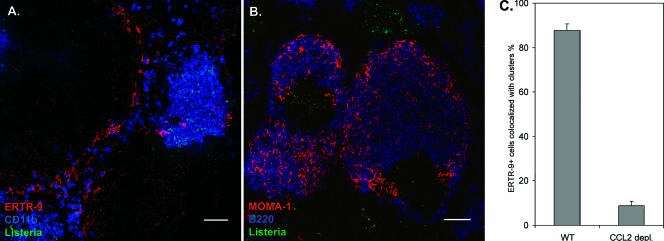
Clustering of phagocytic cells by L. monocytogenes is changed after CCL2 depletion. Spleens were isolated from infected animals treated or untreated with anti-CCL2 24 h after infection. Cryosections were prepared and stained with appropriate fluorescent Abs. A. Migration of ERTR-9+ macrophages is no longer observed in CCL2-depleted mice, and bacteria are no longer associated with these macrophages. CD11b cells are stained blue, ERTR-9 is red, and Listeria cells are green. B. Migration of MOMA-1+ cells into the B-cell follicle is unaltered in CCL2-depleted mice compared to control BALB/c mice. MOMA-1 cells are stained red, B220 is blue, and Listeria cells are green. Bars, 50 μm. These experiments were done twice using three mice. More than 20 fields of view were analyzed in each experiment. The control staining of spleens from infected but untreated mice is depicted in Fig. 2, since these two experiments were done in parallel. C. Percentage of ERTR-9+ cells associated with infectious foci at 24 h p.i. in BALB/c mice treated with anti-CCL2 compared to untreated control mice. ERTR-9+ cells were counted in fields of view of immunohistological sections from spleens of at least three treated and untreated mice, and means and standard errors were calculated. More than 10 fields of view were analyzed for each condition.

