FIG. 2.
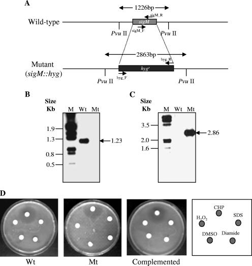
Strategy for construction of sigM deletion and ΔsigM complementation strains and the resulting effects on the growth phenotype of M. tuberculosis CDC1551. (A) The M. tuberculosis sigM locus and strategy for deletion mutant construction. (B and C) Southern blot analysis of wild-type M. tuberculosis CDC1551 (Wt) and sigM deletion mutant (Mt). Genomic DNA of each strain was digested with PvuII, electrophoresed in a 0.8% agarose gel, and transferred to a nylon membrane. The resulting blot was UV cross-linked and subsequently probed with either the sigM-specific PCR amplicon or a hyg gene-specific PCR amplicon. The sigM-specific probe was prepared by amplifying the DNA fragment using the primers sigM_F (5′-GCAGCGGGCACTGATGCG-3′ and sigM_R (5′-TAGCCCAGCAGCCGCGC-3′) and digoxigenin-labeled dUTP (Roche); similarly, the hyg gene-specific probe was prepared by amplifying the DNA fragment using the primers hyg_F (5′-GGGAATTCCATATGACACAAGAATCCCTG-3′) and hyg_R (5′-CCTTAATTAATCAGGCGCCGGGGGCGGT-3′) and digoxigenin-labeled dUTP (Roche). Expected sizes for the PvuII fragments from the wild-type and sigM mutant strains were 1.2 and 2.9 kb, respectively. (D) Susceptibility testing of different mycobacterial strains to various oxidative stresses and detergent stress (each 10 μl) by disk diffusion assay using 4.5-mm Whatman paper disks. The photographs demonstrate the effects of the oxidative stresses, cumene hydroperoxide (20 mM), diamide (1 M), and hydrogen peroxide (H2O2; 100 mM), and the detergent, SDS (1%), on the survival of wild-type (Wt), ΔsigM mutant (Mt), and ΔsigM-complemented strains of M. tuberculosis. The effects of these stress agents were determined by measuring the diameter of the zone of inhibition. These photographs were obtained with three individual sets of experiments and a representative photograph is shown in the figure. Since the cumene hydroperoxide is prepared as a DMSO solution, a negative control with 10 μl of DMSO was used.

