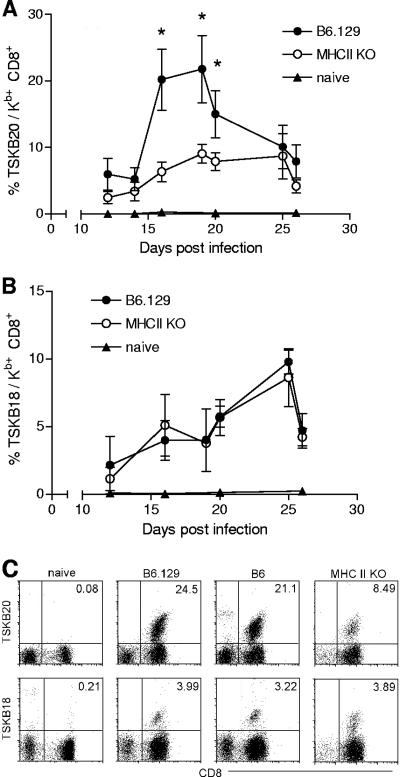
FIG. 1.
Antigen-specific CD8+ T cells develop in the absence of CD4+ T-cell help. Blood from uninfected naïve mice (filled triangles), infected B6.129 wild-type mice (filled circles), and infected MHC II KO mice (empty circles) was sampled at the indicated time points after infection and stained with H-2Kb tetramers bearing the TSKB20 (A) or TSKB18 (B) T. cruzi peptide. Values represent the means ± SD of tetramer-positive cells among CD8+ T cells. Statistically significant differences (P < 0.05) in the frequencies of peptide-specific CD8+ T cells between MHC II KO and wild-type mice are indicated by asterisks. (C) TSKB20/Kb and TSKB18/Kb staining of blood from naïve mice, infected B6.129 mice, infected B6 mice, and infected MHC II KO mice at 19 days postinfection. Cells shown are gated on CD4− CD11b− B220− lymphocytes. Data are representative of two experiments (n = 3 to 5 mice per group).

