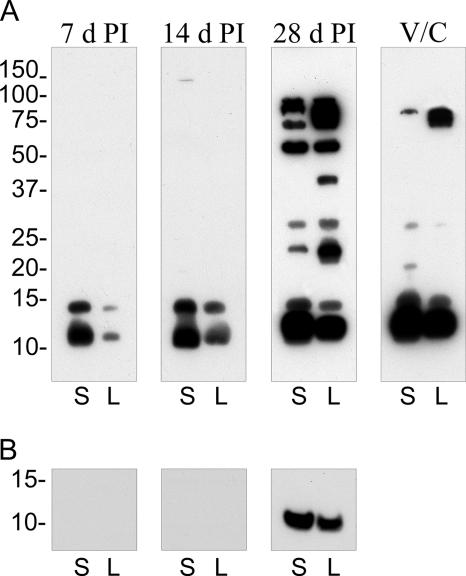
FIG. 3.
Temporal analysis of antibody development against SCV and LCV antigens. (A) Immunoblots containing an equal amount of SCV (S) and LCV (L) lysates were probed with sera derived from a C. burnetii-infected guinea pig at 7, 14, and 28 days p.i. or with serum derived from a guinea pig vaccinated with formalin-fixed C. burnetii and then challenged with live organisms (V/C) (see Materials and Methods). (B) Immunoblot of proteinase K-digested lysates probed with infected guinea pig sera. Depicted is the region of the immunoblot showing a digestion-resistant product that comigrates with Nine Mile Crazy LPS. Film exposures for all immunoblots were conducted for 30 s. Molecular mass markers at left are expressed in kDa.

