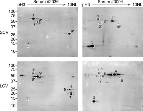
FIG. 4.
Two-dimensional immunoblots of SCV and LCV lysates showing antigens recognized by convalescent-phase sera from two human patients who had recovered from Q fever. Isoelectric focusing of proteins was conducted using pH 3 to 10 nonlinear (NL) strips. Immunoreactive spots were cross-referenced with parallel silver-stained gels. Stained proteins that clearly correlated with immunoreactive spots were processed for mass spectrometry. Identified antigens are denoted with circles and numbers. Spots on other gels that have coordinates identical to those of mass spectrometry-identified antigens are denoted with circles and asterisked numbers. Identified antigens are listed in Table 2. (Some immunoreactive protein spots are difficult to visualize in the figure but are clearly visible in the original films.) Molecular mass markers at left are expressed in kDa.

