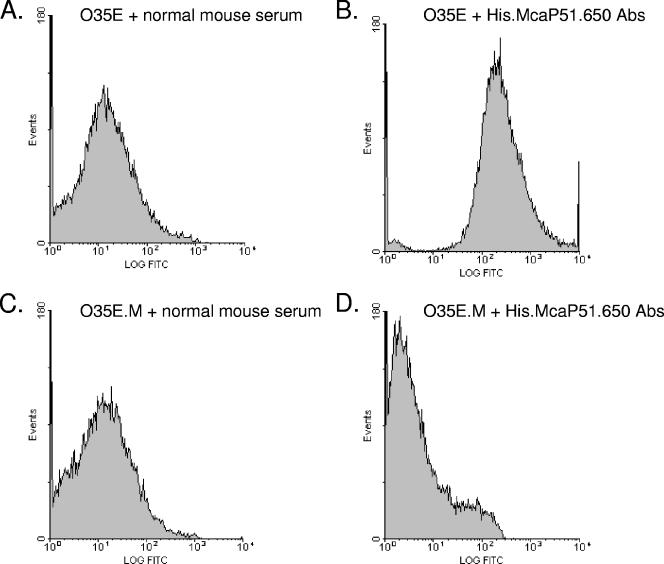
FIG. 3.
Flow cytometry analysis of M. catarrhalis strains O35E and O35E.M. M. catarrhalis cells were incubated with normal mouse serum (A and C) or murine serum containing His.McaP51.650 antibodies (Abs) (B and D) at a dilution of 1:25. Bacteria were washed, incubated with FITC-conjugated secondary antibody, and processed as described in Materials and Methods. The x axes represent the level of fluorescence, and the y axes correspond to the particles counted in arbitrary units.

