FIG. 3.

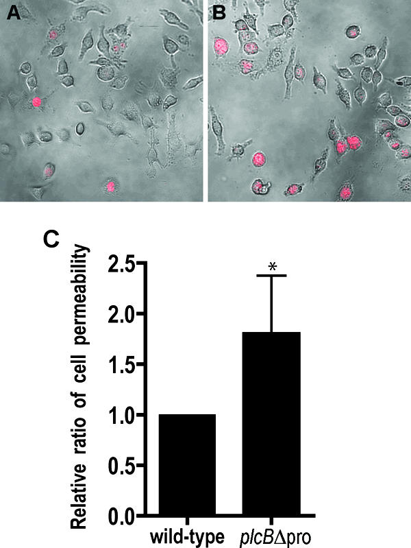
Staining of live infected J774 cells with a fluorescent, non-membrane-permeant nucleic acid dye. Host cells were infected with the wild type or the plcBΔpro mutant for 6 h, with the addition of 50 μg/ml gentamicin between 1 h and 3 h postinfection. Live infected cells were stained at 6 h postinfection. Images were captured with a ×40 lens, using a confocal microscope with a krypton laser and transmitted light. Host cells with compromised membranes are stained red. The images show overlays of phase-contrast and fluorescence images of host cells infected with the wild-type strain (A) or the plcBΔpro mutant (B). (C) Ratios of stained cells to the total number of infected host cells (1 × 10³ to 2 × 10³ per sample; n = 5 per sample). The wild type was set to have a ratio of 1. Statistical analysis of the data indicated that the P value was 0.018.
