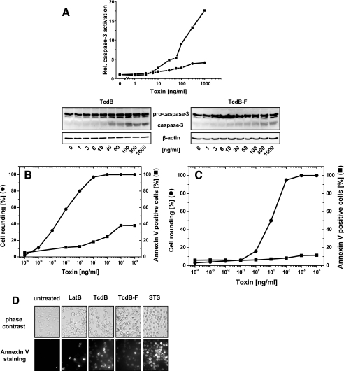
FIG. 2.
Cytotoxic effect in nonsynchronized RBL cells. (A) Activation of caspase-3 by TcdB. RBL cells were exposed to increasing concentrations of TcdB (▪) or TcdBF (•) for 3 h. The cellular levels of procaspase-3 and caspase-3 were determined by Western blot analysis. The signal intensity was normalized to the intensity of the beta-actin signal. (B) Cytotoxic effect of TcdB. RBL cells were exposed to increasing concentrations of TcdB for 24 h. The cytopathic effect was determined as the percent rounded cells. Annexin V-positive cells were stained with annexin V-Alexa Fluor 488 and visualized by fluorescence microscopy. (C) Cytotoxic effect of TcdB. RBL cells were exposed to increasing concentrations of TcdBF for 24 h. The cytopathic effect was determined as the percent rounded cells. Annexin V-positive cells were stained with annexin V-Alexa Fluor 488 and visualized by fluorescence microscopy. (D) Cytotoxic effect in nonsynchronized RBL cells. RBL cells were exposed to latrunculin B (2.5 μM), staurosporine (STS) (0.2 μM), TcdB (0.1 μg/ml), or TcdBF (1 μg/ml) or left untreated for 24 h. Phosphatidylserine exposure was visualized by annexin V staining.

