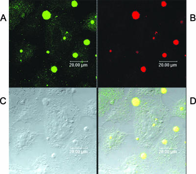
FIG. 9.
Confocal microscopy of gonococcal microcolonies growing on HEC-1-B cells (C) and stained with human paraglobosyl LOS (1291) IgG F(ab′)2 fragments (red) (B) or stained with MAb 1B2 (green) (A). Panel D is a composite of panels A and B; overlapping binding of the two antibodies is indicated by yellow dots. MAb 1B2, which was induced by human paraglobosyl structures and binds human and neisserial paraglobosyl glycolipids, bound to HEC-1-B cells and bacteria (A and D), while 1291 LOS IgG F(ab′)2 fragments bound to the microcolonies but not to the HEC-1-B cells (B and D).

