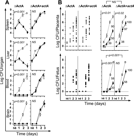
FIG. 3.
Role of ActA in the crossing of the fetoplacental barrier. Pregnant BALB/c mice were injected intravenously at day 14 of gestation with 5 × 10⁵ L. monocytogenes actA deletion mutant cells (ΔActA) or the ΔActA mutant complemented with actA (ΔActA+actA). As compared to the isogenic mutants, bacterial growth for the EGDwt strain is indicated by the dotted lines. Animals were monitored by quantifying bacterial growth at the indicated times. (A) Bacterial growth in organs (spleen, liver, and brain) and in blood of pregnant mice infected with ΔActA (left panels) and actA-complemented ΔActA+actA (right panels) strains. Results shown are means ± standard errors from groups of three to five mice and are expressed as the log10 bacteria (CFU) per organ or log10 bacteria per ml of blood. (B) The kinetics of bacterial growth are represented on the left panels by dot plots corresponding to individual bacterial counts for each placenta (top panel) and each fetus (lower panel) for the indicated times and strains. In the right panels, the means ± standard errors for each time and strain are given for ΔActA and the actA-complemented ΔAct mutant and P values for the comparison between the two strains are also reported. The numbers reported near each value correspond to the percentage of infected placenta among all analyzed placentas. All data are representative of three independent experiments. P values reported in each graph correspond to the comparison between EGDwt and the indicated mutant. NS, not statistically different.

