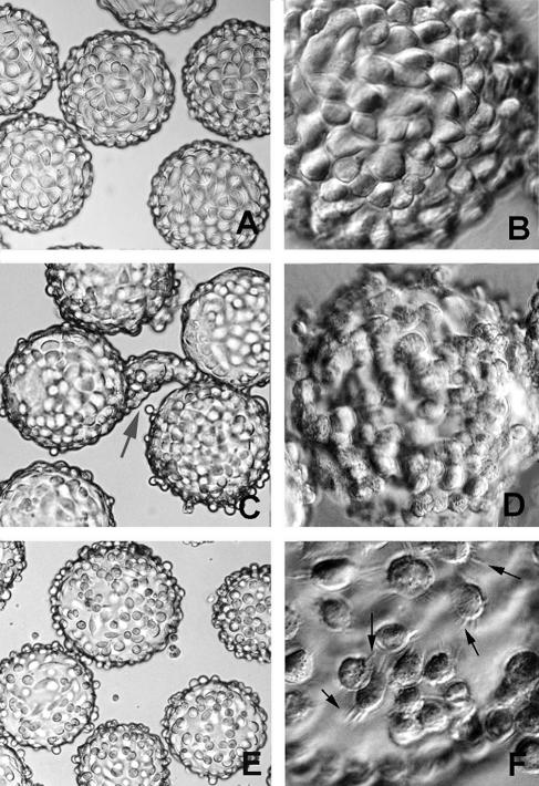
FIG. 3.
Light microscopy analysis of McCoy, HEC-1B, and HeLa cell monolayer formation on microcarrier beads. (A and B) Even distribution of McCoy cells over the bead surface, 2 days after initial seeding. (C and D) Confluent HEC-1B monolayers were obtained 7 to 10 days after seeding. Polarized endometrial cells tend to form small and large “organoid”-like structures (gray arrow in panel C); with time, the organoids detach, leaving a lush monolayer (D). (E) Uneven distribution of HeLa cells over the bead surface, with the presence of free-floating cells at day 3 after seeding. (F) Extensions emanate from the HeLa cells (black arrows) as if trying to anchor to the collagen surface, but spaces remain. Magnifications: ×150 (A, C, and E) and ×450 (B, D, and F).

