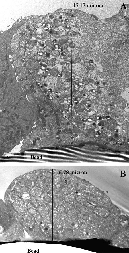
FIG. 6.
Transmission electron photomicrograph sagittal views of HEC-1B and HeLa epithelial cells from bead cultures infected with C. trachomatis serovar E for 36 hpi. Epithelial cells grown on beads established monolayers with architectural characteristics similar to the tissue from which they originated; the tall columnar endometrium-derived HEC-1B cells on beads (A) were, on average, two- to threefold taller than the low columnar cervical-derived HeLa cells grown in the same conditions (B). Consistent with other data, chlamydial inclusions were larger and more mature in HEC-1B (A) than in HeLa (B) cells. Magnification: ×7,530.

