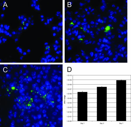
FIG. 2.
F. tularensis localizes to the alveolus following inhalation. Mice were intranasally inoculated with 105 CFU of F. tularensis LVS expressing GFP. One, three, and seven days postinoculation, lungs were harvested and prepared for immunofluorescence analysis. (A to C) Bacterial localization was determined by probing lung sections with a fluorescently labeled antibody to GFP (green). Nuclei were stained with DAPI (blue) to visualize lung cells. Representative images of the alveoli of infected mice 1 (A), 3 (B), and 7 (C) days postinoculation. (D) Bacterial recovery from lungs 1, 3, and 7 days following intranasal inoculation with 105 CFU LVS. Each bar represents mean recovery from three mice; error bars represent standard deviations of the means.

