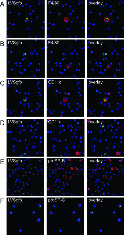
FIG. 4.
F. tularensis LVS expressing GFP colocalized with cells expressing the macrophage marker F4/80, the dendritic cell marker CD11c, and the ATII cell markers proSP-B and proSP-C. Nuclei were stained with DAPI (blue). Mouse lung cells were probed with fluorescently labeled antibody to F4/80 (red) (A and B), CD11c (red) (C and D), proSP-B (red) (E), and proSP-C (red) (F). Representative images are from lung cells 3 days postinoculation.

