Figure 5.
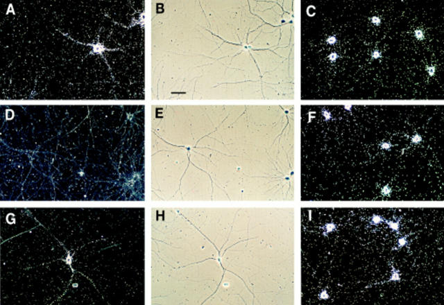
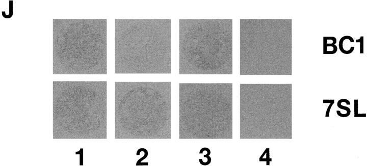
Regulation of BC1 expression by neuronal activity. Hippocampal neurons were grown in culture for 14 d in the absence of TTX (A–C), in the presence of 1 μM TTX (D–F), or in the presence of 1 μM TTX for the first 9 d, and in the absence of TTX for the following 5 d (G–I). Left (A–G): BC1 RNA, dark-field photomicrographs; middle (B–H), BC1 RNA, phase contrast photomicrographs, corresponding to dark-field photomicrographs in left column; right (C–I): 7SL RNA, dark-field photomicrographs. Dark field photomicrograph D (BC1 RNA in the presence of TTX) was overexposed to reveal absence of any significant labeling over cells or neurites. All cultures were grown at medium density. Bar, 50 μm. (J) Cultured hippocampal neurons on coverslips were exposed to autoradiographic film. Autoradiographs show BC1- and 7SL-labeling signals for cells that were grown for 14 d in the absence of TTX (1), in the presence of 1 μM TTX (2), or in the presence of 1 μM TTX for the first 9 d and in the absence of TTX for the following 5 d (3). (4) Sense strand control.


