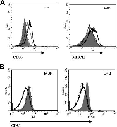
FIG. 1.
DC activation by MBP. (A) DCs were treated with 10 ng/ml (dotted line), 100 ng/ml (thin black line), or 1 μg/ml (thick black line) of MBP for 24 h, and surface expression changes of CD80 (left) and MHC-II (right) were compared to those of untreated controls (solid histograms). (B) DCs were exposed to 10 μg/ml of MBP (left) or 1 μg/ml of LPS (right) for 24 h in the presence (dotted line) or absence (solid histogram) of 5 μg/ml of polymyxin B. Changes of CD80 surface expression were compared to those of untreated controls (solid black line). The data are representative of two independent experiments.

