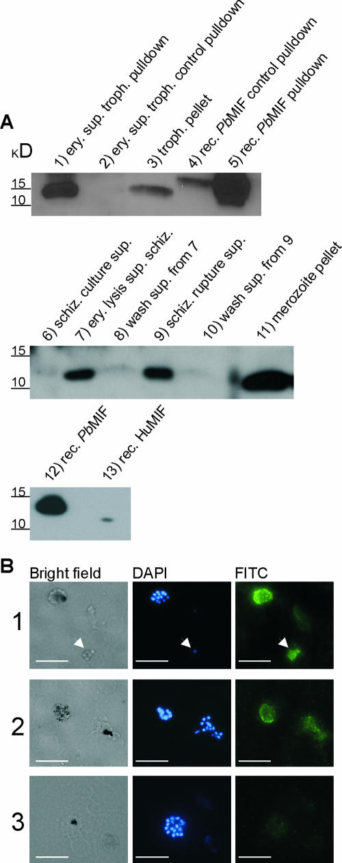
FIG. 4.
PbMIF is externalized by P. berghei and is released upon schizont rupture. (A) Western blot analysis using anti-PbMIF rabbit polyclonal antiserum. Lane 1 contained erythrocytes (ery.) infected with trophozoite (troph.) stage P. berghei that were isolated by heart puncture and lysed by osmotic shock. Parasite-derived MIF was pulled down from the lysis supernatant (sup.) (from the equivalent of 107 infected erythrocytes) using anti-PbMIF bound to protein G-Sepharose. Lane 2 contained control pulldown of the lysis supernatant using protein G-Sepharose alone. Lane 3 contained a trophozoite pellet (∼2.0 × 10⁵ parasites loaded). Lane 4 contained control pulldown of 100 ng of recombinant (rec.) C-terminally His6-tagged PbMIF using protein G-Sepharose alone. Lane 5 contained control pulldown of 100 ng of recombinant C-terminally His6-tagged PbMIF using anti-PbMIF loaded protein G-Sepharose. Lane 6 contained supernatant (10 μl of 100 ml) of an overnight schizont (schiz.) culture. Lane 7 contained supernatant (10 μl of 20 ml) of schizonts after disruption of the host erythrocyte membrane. Lane 8 contained phosphate-buffered saline wash supernatant (10 μl of 1 ml) of schizont pellet from lane 7. Lane 9 contained supernatant (10 μl of 1 ml) after mechanical rupturing of the schizonts into merozoites. Lane 10 contained phosphate-buffered saline wash supernatant (10 μl of 1 ml) of the merozoites from lane 9. Lane 11 contained solubilized merozoite pellet (∼2.0 × 10⁵ parasites loaded). Lane 12 contained 50 ng of recombinant C-terminally His6-tagged PbMIF. Lane 13 contained 100 ng of recombinant huMIF. (B) Immunofluorescent detection of c-myc-tagged PbMIF in blood stages. Thin smears from an overnight schizont culture expressing PbMIF-c-myc were stained using an anti-c-myc monoclonal antibody. (Panel 1) Intact schizont and trophozoite (arrowhead) show staining within the parasitophorous vacuole. (Panel 2) Intact schizont and ruptured schizont. (Panel 3) Wild-type P. berghei control stained with the anti-c-myc monoclonal antibody. The fluorescent image in panel 3 was taken with a 0.6-s exposure, while the fluorescent images in panels 1 and 2 were taken with a 0.3-s exposure. Bars, 10 μm. FITC, fluorescein isothiocyanate.

