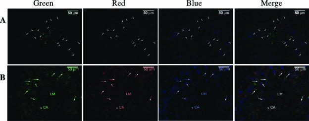
FIG. 4.
NK1.1+ CD11+ cells express IFN-γ in situ. Splenic frozen sections from mouse infected with 4,000 L. monocytogenes organisms 24 h earlier were stained for L. monocytogenes (green), Ly49G2 (red), and CD11c (blue) (A) or IFN-γ (green), Ly49G2 (red), and CD11c (blue) (B). (A) The arrows indicate Ly49G2+ CD11c+ cells. (B) An arrowhead indicates a central arteriole (CA), and the arrows denote the Ly49G2+ CD11c+ IFN-γ+ cells. “LM” indicates the putative infectious focus.

