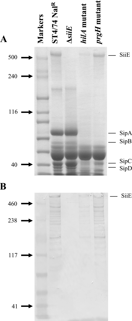
FIG. 2.
The secretion of SiiE is influenced by hilA but not T3SS-1. (A) Proteins isolated from culture supernatants were separated by SDS-PAGE and stained with Coomassie blue. Molecular masses (kilodaltons) of standard proteins are shown on the left. The locations of T3SS-1-secreted proteins (SipA to SipD) are indicated on the right. (B) Proteins isolated from culture supernatants were transferred onto membrane by Western blotting and probed with the anti-SiiE monoclonal antibody. Molecular masses (kilodaltons) of standard proteins are shown on the left. Lanes are as shown for panel A.

