
Fig. 10.
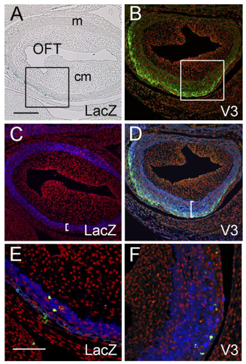
V3 causes an increase in thickness of the myocardium of the proximal cardiac outlet. Adenovirus expressing versican V3 or LacZ were injected into the anterior heart field (AHF) of Hamburger and Hamilton stage (HH) 19 chick embryos and evaluated at HH29. A: X-gal staining (blue) of the proximal outflow tract (OFT) of an embryo injected with LacZ-expressing virus. B: Anti– hyaluronantag staining (green) of the proximal OFT of an embryo injected with V3-expressing virus. C: Anti–α-sarcomeric actin staining (blue) of the same section shown in A demonstrates that LacZ-expressing cells are present within the myocardial sleeve of the OFT. D: Anti–α -sarcomeric actin staining (blue) of the same section shown in B demonstrates that V3-expressing cells are present within the myocardial sleeve of the OFT. Brackets in C and D indicate the thickness of the OFT myocardium. E and F show phosphohistone H3 immunodetection to assess cell proliferation within the OFT myocardium (green). Propidium iodide was used to stain the nuclei of cells (red). Scale bars = 150 μm in A (applies to B–D), 50 μm in E (applies to F).