
Fig. 5.
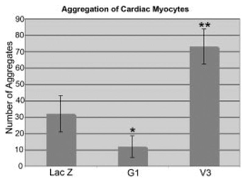
V3 promotes myocardial cell aggregation. Primary cardiac myocyte cultures were infected with adenovirus expressing LacZ, the G1 domain of versican, or the V3 versican variant. Nuclear condensations were counted 72 hr after infection. Values represent the average of three different experiments in which three separate optical fields of cell aggregates were counted. P values were calculated using an analysis of variance test coupled with the conservative Bonferroni’s analysis: the overall test results produced an F = 37.48 with a corresponding P = 0.0000; *pairwise analysis showed G1 and LacZ with a mean difference of 19.5, with a corresponding, P = 0.023, the G1 and V3 mean difference is 60.25 with a corresponding P = 0.000, the **V3 and LacZ mean difference is 40.75, with a corresponding P = 0.000.