
Fig. 9.
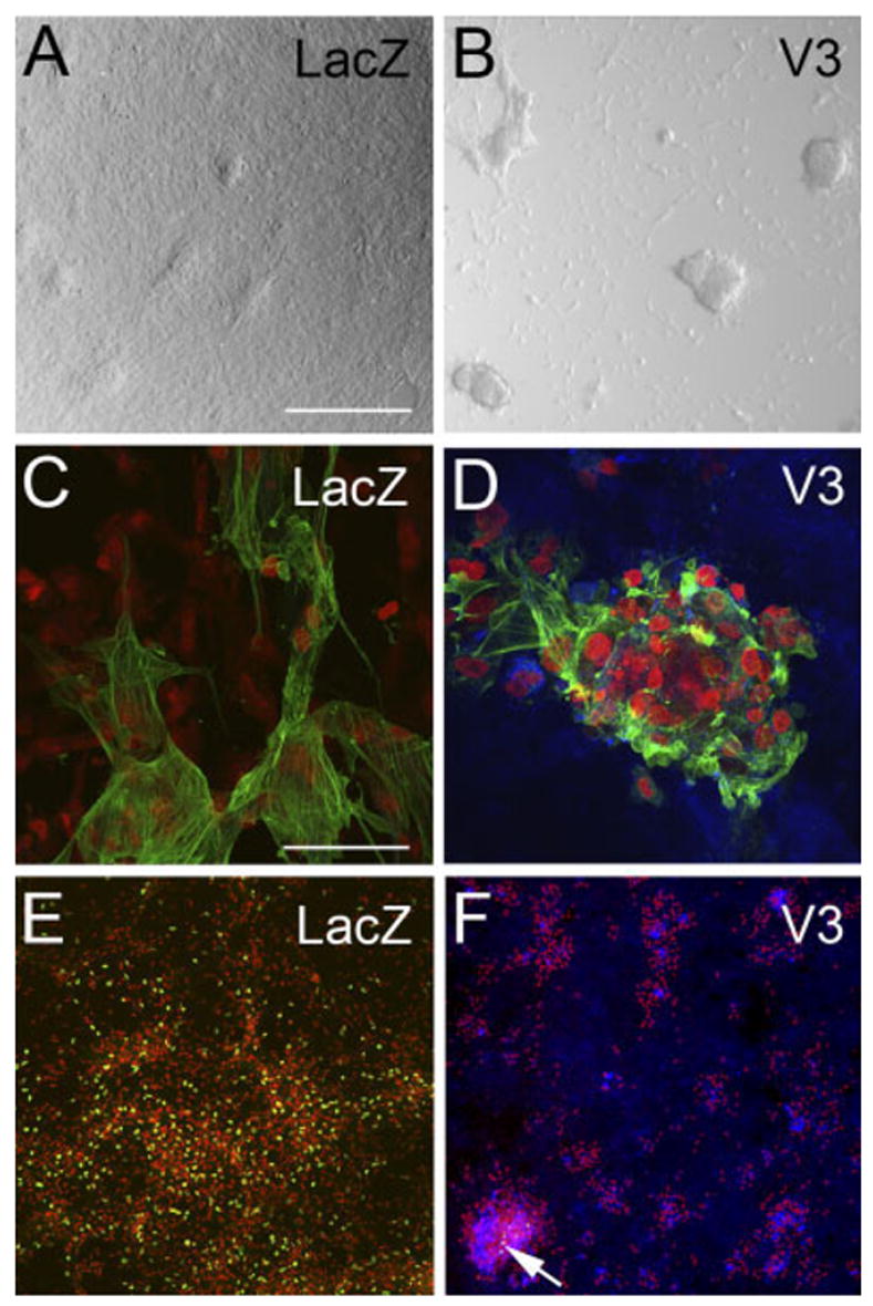
Expression of the noncleavable versican V3 variant in myocardial cells caused an increase in cell– cell aggregation. A,C,E: Primary cardiomyocyte cultures were infected with adenovirus expressing LacZ. B,D,F: Primary cardiomyocyte cultures infected with the versican variant V3. A,B: Cultures infected with V3 developed more cell aggregates (B) than control-infected cultures (A). C,D: Immunodetection using α-sarcomeric actin antibody (green). The results indicate that aggregates were composed of myocardial cells. D,F: Immunolabeling using anti– hyaluronan tag (blue). The results indicate that recombinant V3 is expressed within the aggregates as well as in the extracellular matrix (ECM) of cells throughout the culture. E,F: Bromodeoxyuridine (BrdU) labeling as a measure of cell proliferation (green nuclei). Propidium iodide was used to stain the nuclei of cells (red). Arrow in F shows BrdU-positive cells that represent a very small fraction of total cells in the dish. See Figure 5 for quantification of the myocardial cell aggregates. Scale bars = 300 μm in A (applies to B,E,F), 50 μm in C (applies to D).