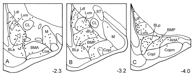
Fig. 1.

Distribution of 5-HT3AR+ neurons in the rat amygdala. Neurons were plotted from two 50 μm thick sections at each level of the amygdala (Bregma −2.3, −3.2, and −4.0 of Paxinos and Watson, 1997). A third section located between the two sections was stained with cresyl violet to identify nuclear borders. Abbreviations: AHA, amygdalohippocampal area; BLa, anterior subdivision of the basolateral nucleus; BLp, posterior subdivision of the basolateral nucleus; BMA, anterior basomedial nucleus; BMP, posterior basomedial nucleus; CL, lateral subdivision of the central nucleus; Coa, anterior cortical nucleus; Copl, posterolateral cortical nucleus; Copm, posteromedial cortical nucleus; Ldl, dorsolateral subdivision of the lateral nucleus; Lvm, ventromedial subdivision of the lateral nucleus; M, medial nucleus; ST, stria terminalis.
