
Figure 3.
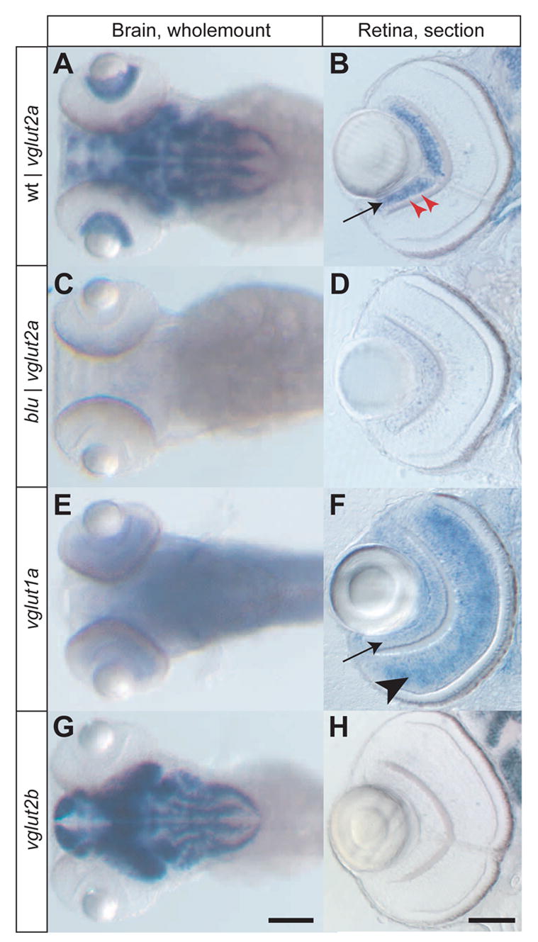
Expression patterns of three zebrafish vglut genes, detected by RNA in situ hybridization. Images in the left column (A, C, E, G) show wholemount stainings of the head at 4 dpf, with antisense probes for the genes indicated. The right column (B, D, F, H) show vibratome sections (20 μm) of zebrafish eyes at 4 dpf. A, B, vglut2a is expressed in a complex pattern in the brain of wildtype and in apparently all RGCs in wildtype (arrow). A single row of GABAergic amacrine cells in the RGC layer is negative for vglut2a (red arrowheads). C, D, vglut2a RNA (containing a translational stop) is absent in both brain and eye of blu mutants. E, F, vglut1a RNA is found in a more diffuse pattern than vglut2a in the brain. In the retina, it is strongly expressed in bipolar cells (arrowhead) and weakly in RGCs (arrow). G, H, vglut2b RNA is strongly expressed in a pattern similar to vglut2a in the brain and is absent from the retina. Scale bar in G: 100 μm (for A, C, E, G); scale bar in H: 50μm (for B, D, F, H).