
Figure 6.
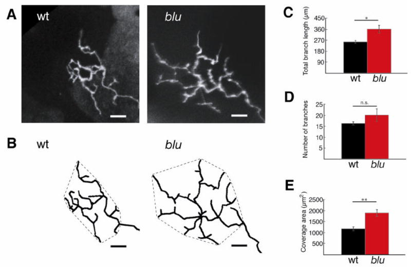
RGC axon arbors are enlarged in blu mutants. A, Confocal projections of GFP-labeled RGC axon arbors imaged in vivo at 7 dpf. B, Arbors were traced in three dimensions from each branchtip to the first branchpoint of the arbor. Tracings were rotated into a plane parallel to the tectal neuropil and projected. Hatched lines around arbors demarcate the coverage area of the arbor. Scale bar: 10 μm. C–E, Morphometric analysis of RGC axon arbor size reveals that total branch length (C), and coverage area (E) are increased in the mutant. The number of branches (D) is not statistically different. *P <0.05, **P <0.01. Wt, n = 22 cells; blu, n = 11 cells.