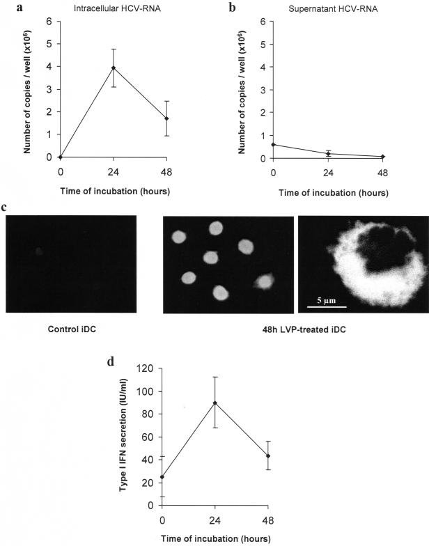
Figure 1. Effect of LVP on immature dendritic cells.
(a, b) iDC were incubated for the indicated times with 6×10⁵ HCV-RNA copies (4 HCV-RNA copies/cell). At d 7, HCV-RNA was extracted from cells (a) and supernatants (b) and quantified. Data represent mean copy number/well±sd from triplicates of one representative experiment out of three. (c) NS5A was detected after cytospin of fixed DC (48 h non-treated or LVP-treated DC) using biotinylated 4F3H2 monoclonal antibody and FITC-conjugated streptavidin. Left: 48 h-non treated DC; middle and right: 48 h LVP-treated DC observed in fluorescence microscopy (middle) and in confocal microscopy (right). (d) Type I IFN production was measured by a biological assay in supernatants of non-treated, 24 h or 48 h LVP-treated DC. Titers are expressed as IU/ml with reference to a recombinant IFN. Data represent mean±sd from four independent experiments.

