
Figure 2.
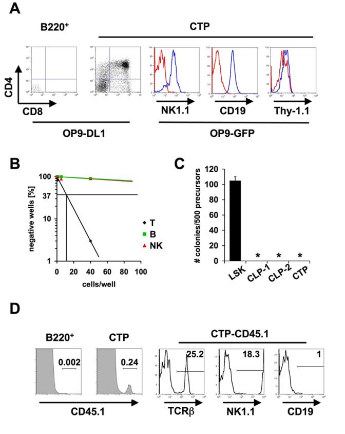
Developmental potential of circulating lin−hCD25+ cells. (A) Sorted lin−hCD25+B220− CTP and lin−hCD25+B220+ cells were co-cultured on OP9-DL1 or OP9-GFP cells for 18 days. Cells were stained for CD4, CD8, CD19, NK1.1 and Thy-1.1 and individual wells were analyzed by FACS. Blue histograms represent specific staining, red histograms represent unstained controls. 800 CD19+ (center) and 550 NK1.1+ (left) cells were recovered from starting cultures of 200 CTP. One representative out of three independent experiments is shown. (B) Limiting dilution analysis of T, B and NK potential of lin−hCD25+B220− cells (CTP) from peripheral blood. 1, 5 or 40 cells were directly sorted onto OP9-DL1 or OP9-GFP cells and analyzed by FACS after 18 days. Wells containing >50 (B and NK potential) or >100 (T potential) lineage positive cells were scored positive. (C) Analysis of myeloid potential of CTP cells. 500 CTP, BM derived LSK, CLP-1 and CLP-2 cells were cultured in methylcellulose containing SCF, IL-3, IL-6 and Erythropoietin and colonies were counted microscopically. (D) 1000 sorted CTP or 50 lin−hCD25+B220+ cells (CD45.1) were injected intravenously into irradiated Rag2−/−γc−/− recipients and spleens were analyzed 5 weeks after transfer by flow cytometry for expression of TCRβ, NK1.1 and CD19. One representative out of 2 independent experiments with 2 mice per group is shown.