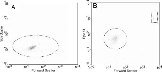
FIG. 2.
Flow cytometric analysis of B. burgdorferi. Wild-type cells were analyzed by flow cytometry in the absence (A) or presence (B) of Syto61. The y axis represents side scatter in panel A and intensity of Syto61 staining in panel B. The dotted circle in panel A indicates that B. burgdorferi cells are detected by side scatter, and that in panel B indicates detection of cells stained with Syto61. Polystyrene beads formed a distinct fluorescent population (dotted box) in the presence of Syto61 (B). Results identical to those in panel B were also obtained with cheA2 mutant cells (not shown).

