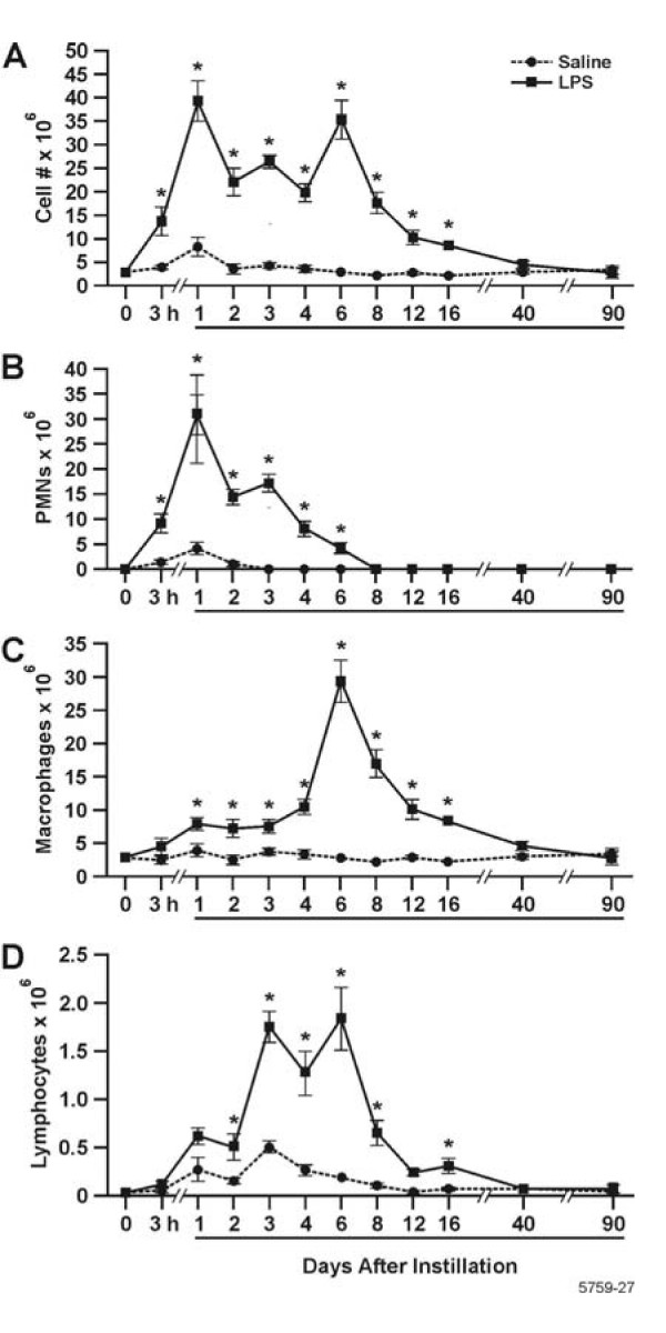
Figure 1.
Three stages of inflammatory cell influx were identified in the BALF, characterized by PMNs, macrophages, and lymphocytes. White blood cells on cytospins were stained with Wright Giemsa and cell counts were performed. A: Total leukocytes; B Neutrophils; C:Macrophages; and D: Lymphocytes. Bars represent group mean values ± SEM (n = 5 rats per experimental group), * = statistically different from saline-instilled controls (P < 0.05).

