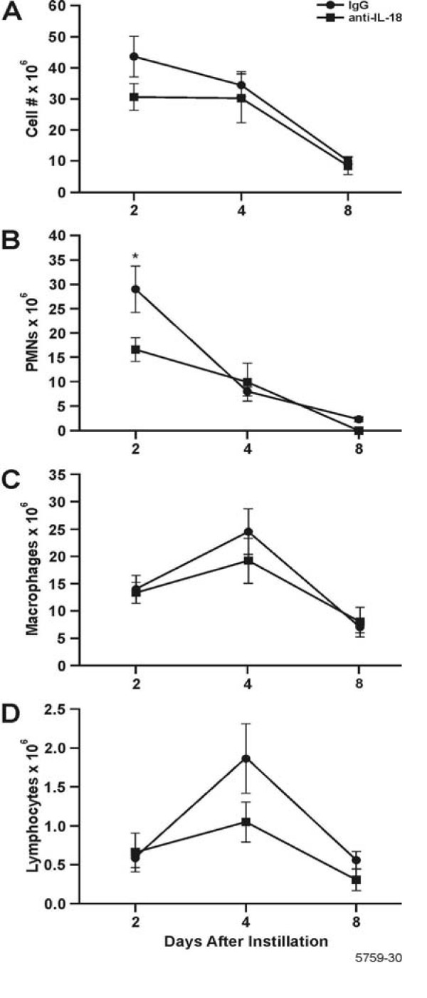
Figure 5.
Inflammatory cell influx in BALF of rats instilled and injected with IL-18 neutralizing antibody and then instilled with LPS. Their lungs were lavaged and cytospin preparations were stained with Wright/Giemsa. The number of (A) Total leukocytes (B) PMNs; * = statistically different from controls treated with IgG1 (P < 0.05). (C) Macrophages;(D) Lymphocytes are shown. n = 3–6 rats per group.

