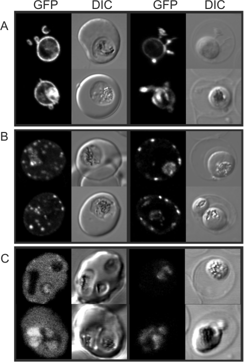
Figure 3. Fluorescence microscopy analysis of the selective permeabilization of host membranes by EqtII.
RBCs infected with (A) EXP1–GFP, (B) Lys119-PfEMP1–GFP, or (C) REX3–GFP transfectants were left untreated (left-hand columns) or treated with 2 HUs of EqtII (right-hand columns). The fluorescence and DIC images were collected using a confocal microscope.

