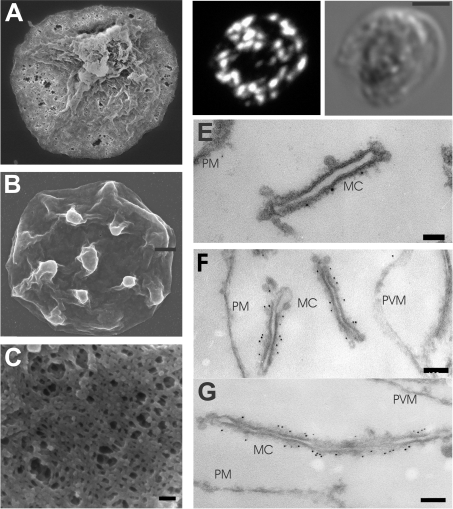
Figure 5. Immunofluorescence and EM analyses of EqtII-permeabilized RBCs.
(A–C) Scanning EM. Isolated mature stage infected RBCs (3D7 strain, 108 cells) were treated with 2 HUs of EqtII (A, C) or mock-treated (B). The cells were pelleted, fixed in 2% (v/v) glutaraldehyde and prepared for scanning EM. (D) Immunofluorescence labelling. Infected RBCs were fixed with 2% paraformaldehyde, then treated with 2 HUs of EqtII, and labelled with anti-PfEMP1 serum followed by Alexa Fluor® 568 anti-rabbit IgG. (E–G) Immuno-EM. Paraformaldehyde-fixed, EqtII-permeabilized PfEMP1–GFP transfectants were labelled with anti-PfEMP1 (E) serum or anti-GFP serum (F, G), followed by Protein A–gold (6 nm conjugate). Abbreviations: MC, Maurer's clefts; PM, RBC plasma membrane; PVM, PV membrane. Scale bars: (A, B) 1 μm; (C) 100 nm; (D) 2.5 μm; (E) 50 nm; and (F) 100 nm.

