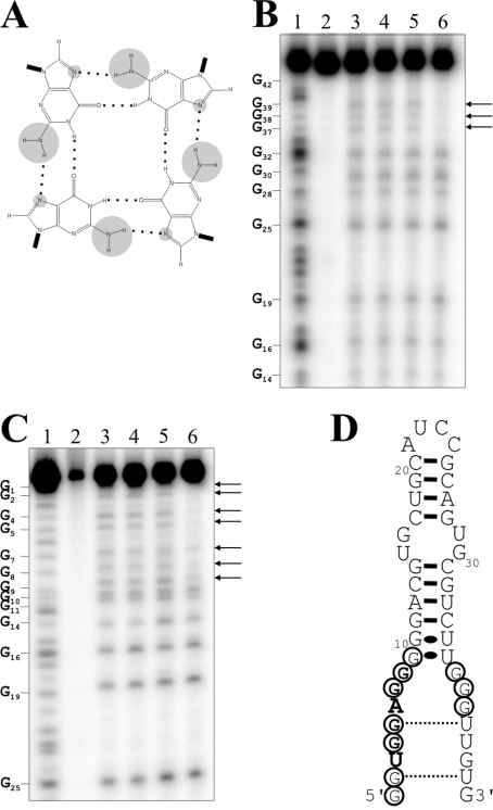
Figure 5. Investigation of a G-quartet-like structure.
(A) Schematic representation of a G-quartet. The hydrogen bonds between the guanosine residues are indicated by dotted lines. The NH2 groups linked to the C2 and the N7 positions are circled in grey. (B) and (C) are autoradiograms of DMS probing of 5′- and 3′-32P-end-labelled ApT4-A' aptamers respectively. Lanes 1: alkaline hydrolyses; lanes 2: the experiments were performed in absence of salt and DMS. Lanes 3–6: show DMS treatments performed either in the absence of salt or in the presence of LiCl, NaCl or KCl respectively. The arrows indicate the resistant guanosine residues. The positions of the guanosine residues are indicated beside the gels. (D) Secondary structure and nucleotide sequence of ApT4-A'. The resistant guanosine residues are circled.

