
Fig. 1.
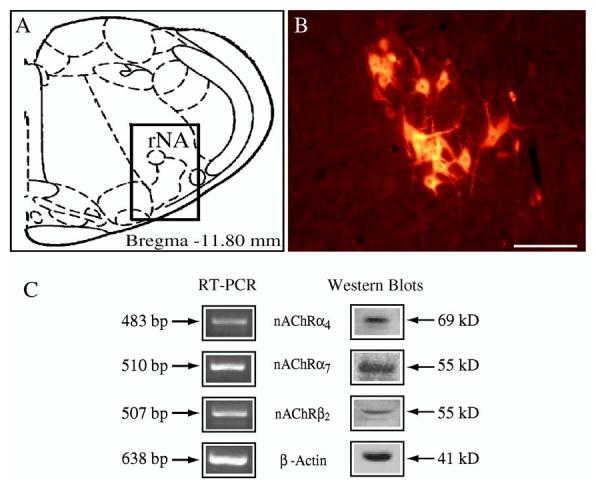
RT-PCR and Western blot analysis of nicotinic acetylcholine receptor mRNA and protein expression in the rostral nucleus ambiguus (rNA) where airway-related vagal preganglionic neurons are located. (A) Schematic representation of a coronal section (Paxinos and Watson, 1986) and (B) the distribution of CTb-labeled AVPNs visualized with rhodamine indicating the location of AVPN within the rNA regions (C), RT-PCR analysis of nicotinic acetylcholine receptor (nAChR) subunits: α4, α7, β2, and β-actin seen at 483, 510, 507, and 638bp, respectively and Western blot analysis of α4, α7, β2, and β-actin, seen at 69, 55, 55, and 41kDa, respectively (Scale bar: 100μm).