
Fig. 3.
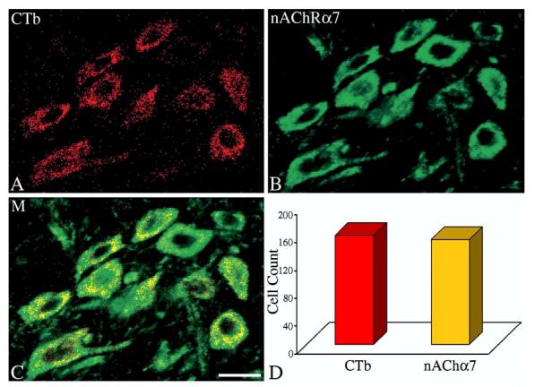
Confocal images of (A), retrogradely labeled airway-related vagal preganglionic neurons (AVPNs) within the rNA, following microinjection of cholera toxin β subunit (CTb) tracer into the upper right lung lobe; (B) α7-nAChR subunit immunoreactive neurons in the same field. (C) The merged (M) image showing the CTb colocalized with an α7-nAChR traits. (D) Total number of counted CTb labeled neurons and the number of the double labeled cells (CTb and α7-nAChR). Scale bar: 20μm.