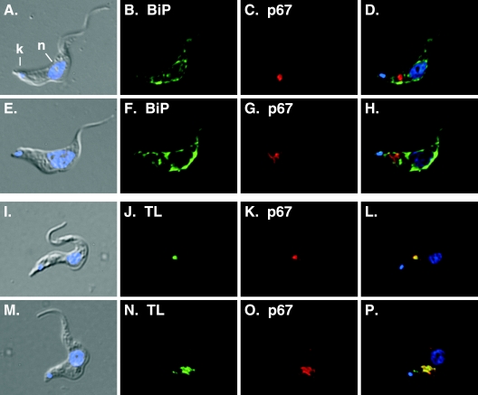
FIG. 4.
Myriocin disrupts lysosomal morphology in trypanosomes. Bloodstream-form trypanosomes were cultured for 14 h in the presence or absence of myriocin (200 nM). Fixed/permeabilized control (A to D) and myriocin-treated (14 h) (E to H) cells were stained with anti-BiP as an ER marker (green) (B and F) or anti-p67 as a lysosomal marker (red) (C and G) and were visualized by epifluorescence microscopy. Alternatively, control (I to L) and myriocin-treated (M to P) cells were allowed to endocytose FITC-TL (J and N) for 1 h and were then processed for staining with anti-p67 (K and O). (A, E, I, and M) Merged DIC and DAPI images; (D, H, L, and P) merged three-channel fluorescent images. DAPI staining (blue) reveals nucleus (n) and kinetoplast (k) localization (indicated in panel A only).

