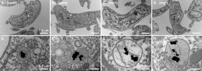
FIG. 5.
Ultrastructure of myriocin-treated trypanosomes. Cultured bloodstream cells were first pulse-loaded (2 h, 37°C) with colloidal gold (5 nm) coupled to bovine holotransferrin to label the terminal lysosome. Cells were then cultured in the absence (A and B) or presence (C and D) of myriocin (200 nM) for 14 h and processed as described for electron microscopy. (A to D) Representative cell sections. Bar, 1.0 μm. White arrowheads indicate lysosomes containing colloidal gold particles. cont, control; myr, myriocin. (E to H) Corresponding images in the lysosomal region taken at a higher magnification. Bar, 0.2 μm.

