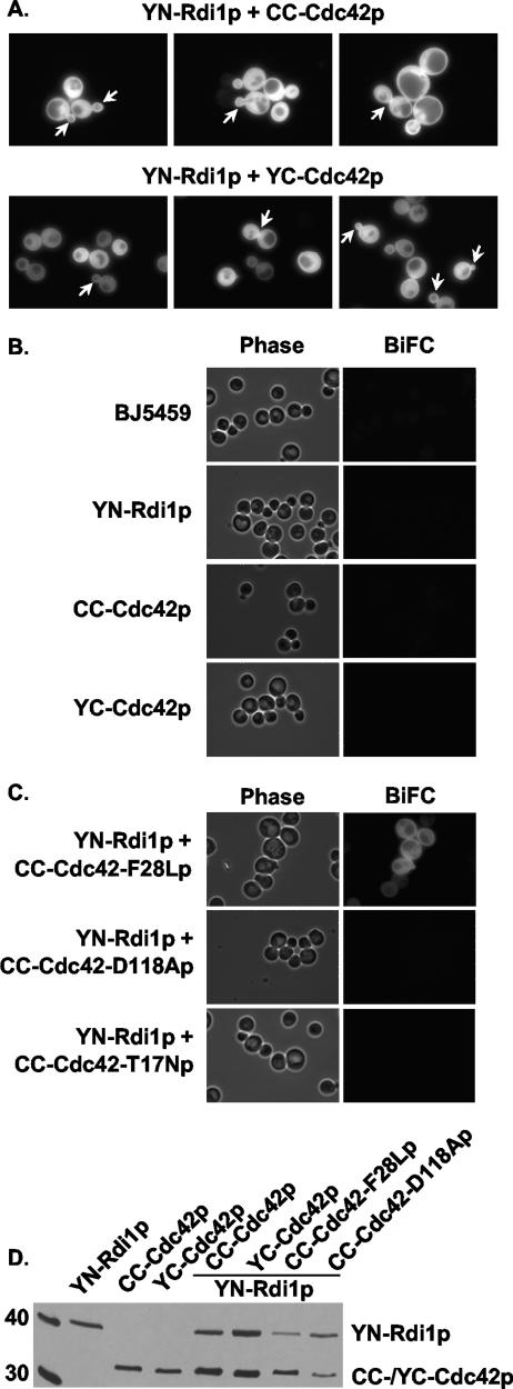
FIG. 1.
BiFC interactions between Cdc42p and Rdi1p. (A) BJ5459 cells expressing YN-Rdi1p and either CC-Cdc42p or YC-Cdc42p were grown in low-fluorescent SC medium without Met to mid-log phase and observed by fluorescence microscopy. Arrows indicate an enhanced BiFC signal at the tips and sides of small- and medium-sized buds and at the mother-bud neck region. (B) BJ5459 cells expressing either YN-Rdi1p, CC-Cdc42p, or YC-Cdc42p alone were grown and observed as described above (A). (C) BJ5459 cells expressing YN-Rdi1p and either CC-Cdc42(F28Lp), CC-Cdc42(D118Ap), or CC-Cdc42(T17Np) were observed as described above (A). (D) Immunoblot analysis of Cdc42p and Rdi1p fusion proteins. Thirty micrograms of total protein from BJ5459 cells expressing the indicated proteins were resolved on 13% SDS-PAGE gels and immunoblotted with anti-GFP α-Av antibody. Left lane, size markers (in kDa).

