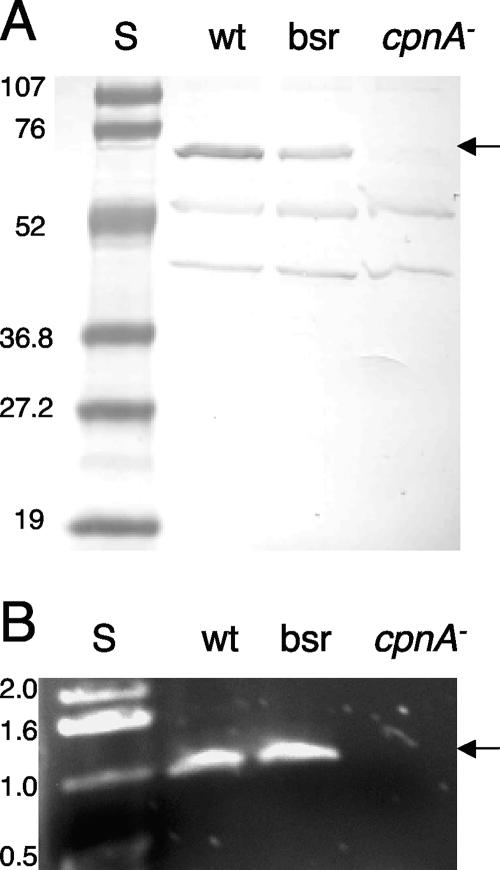
FIG. 1.
Analysis of cpnA− null mutants. (A) Whole-cell samples (5 × 10⁶ cells per lane) from wild-type (wt) cells, nonhomologous recombination blasticidin-resistant (bsr) control cells, and cpnA− cells were analyzed by Western blotting with CpnA antiserum. Protein standards (S) are in the first lane, with molecular masses indicated in kDa. The arrow points to the CpnA protein band. (B) Wild-type cells, nonhomologous recombination blasticidin-resistant control cells, and cpnA− cells were lysed and analyzed by PCR using primers that amplify ∼1 kb of the cpnA gene. DNA standards are in the first lane, with lengths indicated in kb. The arrow points to the amplified cpnA PCR product. Both the CpnA protein and the cpnA gene are absent from cpnA− cells.

