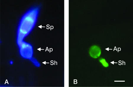
FIG. 5.
Effect of hyperosmotic solute (0.4 M NaCl) on ACE1 expression in a buf1::hph mutant. Spores from a P1.2 buf1::hph transformant expressing promACE1::eGFP vector were inoculated on barley leaves. After 6 h, water droplets were replaced by 0.4 M NaCl and leaves were observed under a microscope 24 h after inoculation. (A) Microscopic observation was performed at ×100 magnification under UV light after staining with calcofluor. A spore (Sp), an appressorium (Ap), and a secondary hypha (Sh) originating from the appressorium are visible through the bright blue fluorescence of their cell walls. (B) Microscopic observation was performed at ×100 magnification under UV light with an eGFP-specific filter. eGFP fluorescence was detected only in the appressorium (Ap) and in the secondary hypha (Sh). Bar, 10 μm.

