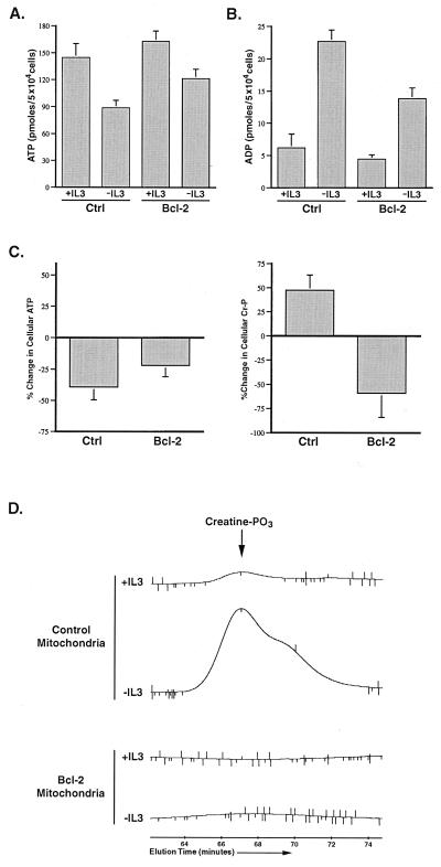
Figure 5.
Growth factor withdrawal in Baf3 cells results in mitochondrial creatine phosphate accumulation that is prevented by Bcl-2 expression. (A) Control (Ctrl) and Bcl-2-transfected Baf3 cells were cultured in the presence (+IL3) or absence (−IL3) of IL-3 for 12 h before the determination of cellular ATP. The data shown are the mean (+SEM) of four independent experiments. The fall in ATP observed on IL-3 withdrawal was statistically significant by Student's t test (P < 0.05). (B) Control (Ctrl) and Bcl-2-transfected cells were cultured in the presence (+IL3) or absence (−IL3) of IL-3 for 12 h, and the amount of cellular ADP present was determined. The data shown are the mean (+SEM) of four independent measurements. The increase in ADP on IL-3 withdrawal was statistically significant by Student's t test (P < 0.05). (C) Control and Bcl-2-transfected cells were cultured in the presence or absence of IL-3 for 12 h before the determination of ATP and phosphocreatine. The percent change in cellular ATP and creatine phosphate (Cr-P) measured on IL-3 withdrawal for each population is shown. The data presented are the mean (+SEM) of four independent determinations. The changes in ATP and phosphocreatine observed in both cases were statistically significant by Student's t test (P < 0.05). (D) Control and Bcl-2-transfected cells were cultured in the presence or absence of IL-3 for 12 h, and perchloric acid extracts normalized to mitochondrial protein were prepared. The amount of creatine phosphate (creatine-PO3) present in the mitochondrial extracts was determined by HPLC. The peak corresponding to the amount of creatine-PO3 present in the mitochondria under each condition is shown. The creatine-PO3 peak in the samples was confirmed by coinjection of purified creatine-PO3 (not shown).

