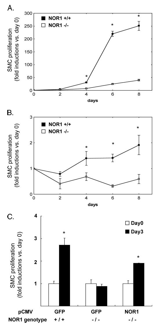
FIGURE 7. NOR1 expression is required for SMC proliferation.

Mouse aortic SMC were isolated from littermate wild type mice and NOR1-deficient mice. Equal numbers of cells (0.5 × 10⁵ cells/plate) were plated on 60-mm plates. A, SMC were maintained in DMEM supplemented with 10% FBS. After 2, 4, 6, and 8 days, the cells were harvested, and cell proliferation was analyzed by cell counting using a hemocytometer. Cell proliferation was expressed as fold induction compared with day 0 and presented as mean ± S.E. (*, p < 0.05 versus NOR1−/−). B, serum-deprived SMC were stimulated with PDGF (25 ng/ml) for the indicated time points, and cell proliferation was analyzed by cell counting. Cell proliferation was expressed as fold induction compared with day 0 and presented as mean ± S.E. (*, p < 0.05 versus NOR1−/−). C, NOR1 wild type or NOR1-deficient SMC were transfected with eukaryotic expression vectors overexpressing either GFP as control or NOR1. Following transfection, cells were maintained in DMEM supplemented with 20% FBS, and cell proliferation was analyzed after 3 days. Proliferation was expressed as fold induction compared with day 0 and presented as mean ± S.E. (*, p < 0.05 versus day 0).
