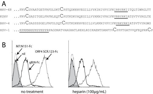
Figure 6. Heparin-independent binding by the N-terminal fragment of gB.
A. Comparison of the gB regions containing the putative heparin binding motif of 3 gamma-herpesviruses-MHV-68, KSHV and BHV-4-and HSV-1. The first 2 conserved cysteine residues of each gB are shown in large type. B. Binding of Fc fusion proteins to NMuMG cells, pre-incubating or not (no treatment) the fusion protein with soluble heparin. gB:N-Fc is the first 423 amino acid residues of gB fused to human IgG1 Fc. 1 of 3 equivalent experiments is shown.

